+91-171-2977793, 3501111-1140 (30 Numbers) info@radicalindia.com, info@radicalscientific.com
Toll Free No.- 1800-1233-229
Confocal Microscopes

RTC-7CON

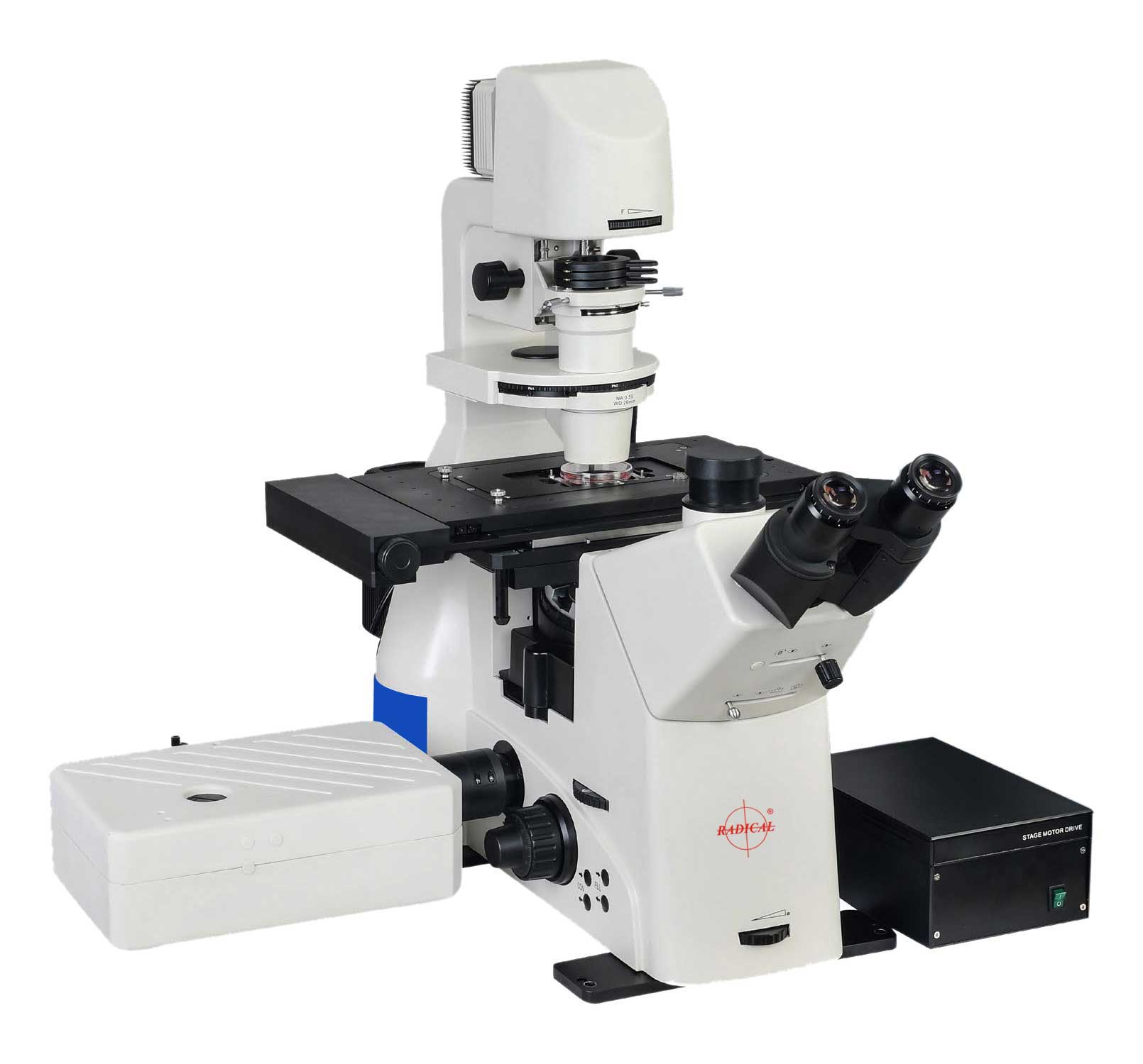
Confocal microscope is a high-end product in Radical Optics microscope series. It is designed as an essential microscopy tool for laboratory scientific research, providing powerful and stable imaging capabilities and highly integrated motorization capabilities.
Efficient scanning head, detector and CVT motorized small hole, coupled with powerful optical system, provides fast, stable, high signal-to-noise (S/N) ratio confocal image.
Confocal Microscope provides a variety of motorized parts, including: motorized stage, motorized focusing, motorized nosepiece, motorized fluorescent carousel, motorized condenser and motorized brightness adjustment, operation mode allows physical button operation and software operation, and provides calling commands, which is convenient for users to control and develop by themselves.

  • High resolution images can be generated with a single click operation, The software will automatically calculate size of the small hole according to objective numerical aperture, exposure value and scanning range, so as to obtain the image with the optimum signal-to noise(S/N) ratio.
  • At same time, noise reduction algorithm can remove the background noise in real time and improve image quality. Multi-channel images can be collected and synthesized simultaneously, which is convenient for customers to realize real-time observation of multiple stains.
  • By setting top position, bottom position and movement interval, the Confocal microscope motorized Z axis can realize automatic Z-Stack acquisitionand generate 3D model.
  • Providing various microscope motorized control interfaces: motorized objective carousel, motorized fluorescent filter unit, motorized condenser turntable.
  • Motorized stage control and motorized focusing mechanism could locate the Region of Interest (ROI) immediately through the software and record the position so that the user will be able to return to the recorded position quickly.
  • Part Name Part Code Description
    Body RCBD Rugged, waterproof, ergonomic modular design, multiple camera ports, touch point treated, (rigid and vibration-free) acid resistant texture painted, intensity controller & on/off switch, RL/TL switch, Eco mode*, rubber feet, slot for ana/pol, transmitted diffusion/filters LBD/ND6/ND25, filter holder, collector, scattering filter, automatic re-adjustable illumination intensity etc, inbuilt diaphragm for different contrast methods. In-built power unit for halogen/led, mounted microscopy stand with provision to attach camera in left side/right side port, dual deck facility.
    Optical System RCiOS Universal infinite fly eye lenses optical system, NIS60 (F200) colour corrected, anti-fungal, anti-bacterial coated (multi-layered hard coated), colour coded objectives, precentered, suitable for bright field, phase / fluorescence / DIC / Emboss /Plas DIC / hoffmann contrast/varel contrast inversion contrast, IMC, DF, ICR, POL, UC-3D imaging with glass and plastic dishes etc.
    Eyepiece RCEP1025 Paired, high eye point WF 10X, FOV-25mm, multilayered coated, high guality, good contrast, abrresion free, aplantic, precentered, diopter adjustment on one/both eyepiece (±5mm)
    Illumination RC5WLED 5W LED cool illumination Life 30,000 hrs, with 4500K (white) (Transmitted) colour temperature, equivalent to 100W halogen, suitable for bright field, phase, IMC, hoffmann, DIC/inversion contrast, Emboss etc. flexible/retroverted ensuring large space, transmitted/epi-fluorescent control knobs located on right side of body-easy to operate, flexible with retroverted (tiltable), buttons for transmitted and reflected easy switch with 2 position filter slider
    Viewing Head RCTST45 Trinocular inclined at 45°, sidentopf head, rotatable through 360°, IPD - from 47-78, diopter adjustment on both eyepiece, top & both side port for camera attachment, splitting ratio100(EP):0(camera) /50:50/0(EP): 100(camera) with 0.7x relay lens placed at side of microscope body, builtin bertrand lens.
    Mechnical Stage RCMC High precision XYZ motorized stage, electric carrier: stroke: 130mm x 110mm (table 325 mm x 144 mm) Maximum speed: 25 mm/s; resolution: 0.1 μ m. Replicate accuracy: 3 μ m. Mechanical adjustable sample splint, attachable mechanical stage:130(X) × 85(Y) mm, accepts different types of micro-testplate, tissue culture bottles, glass slide holder & culture flask holder, universal holder for holding different types of plate’s petri dishes, cell culture plates, specimen slides of all sizes (6, 12, 24, 96 well).
    Nosepiece RC-6N Motorized Inward tilted with positive click stop, Precise sextuple revolving on multiple ball bearing, inbuilt DIC slot
    Condenser RABC Motorized condense, long working distance condenser: NA=0.55,WD=26.00mm, with a full set of DIC, PH functions and related accessories for long time culture tracking of live cells.
    Focusing RXF10 Motorized Z-axis focusing mechanism minimum step length set at 10nm adjustment, Focus up 7 down 2, Coarse stroke 2mm per rotation. Fine stroke 0.002mm per rotation, manual and motorized control, minimum stroke 0.01 µm under motivated control.
    Fluorescence Attachment RCFL6LED Motorized LED fluorescence lamp house, life upto 20,000hrs., 6 positions Attachment removable turret structure with high position filter including 1 for brightfiled, centre adjustable filed diaphragm and 5 holes filter slot for reflected illumination, with following filter, easy to change (push & click). Motorized Shutter. Fluorescent filter block: cover with UV to visible light, Easy to replace.
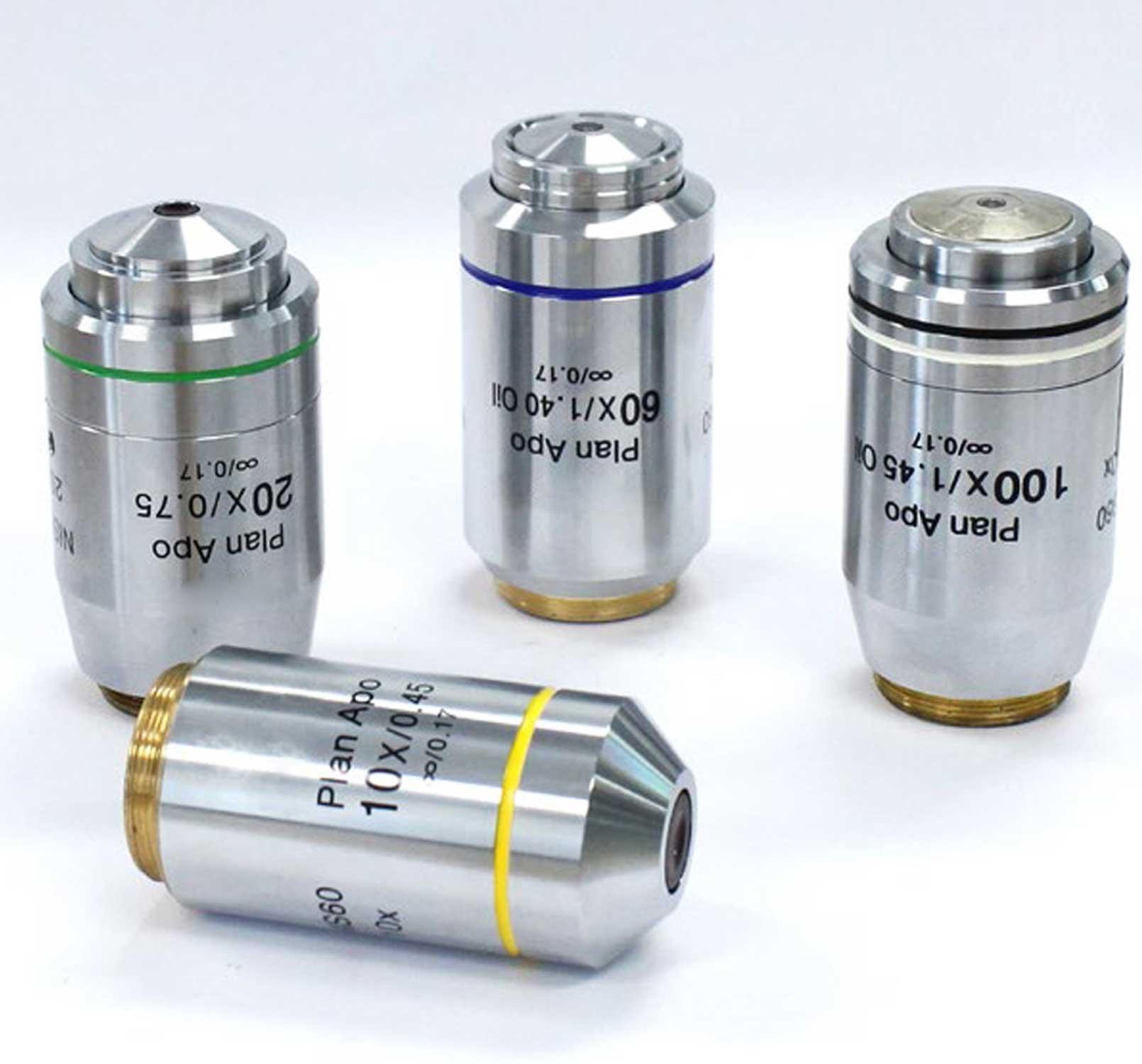
    Objective RC10 10X, NA 0.45, W.D. 4.0mm, cover glass thickness 0.17
      RC20 20X, NA 0.75, W.D. 1.1mm,cover glass thickness 0.17
      RC40 40X, NA 0.95, WD=0.25mm-0.17mm
    Intermediate RCI Manual 1X, 1.5X, Confocal switching
    Output Port RCOP Splitting Ratio: Left: Eyepiece=100:0; Right: Eyepiece=100:0
    DIC Plate RCDP6 10X,20X,40X, Plate, Can be Inserted in Nosepiece Slot, motorized with analyzer & polarizer attachment. Slider and modules for objectives
    Controller RCC Rocking Bar, Controller Box, USB Connection Cable
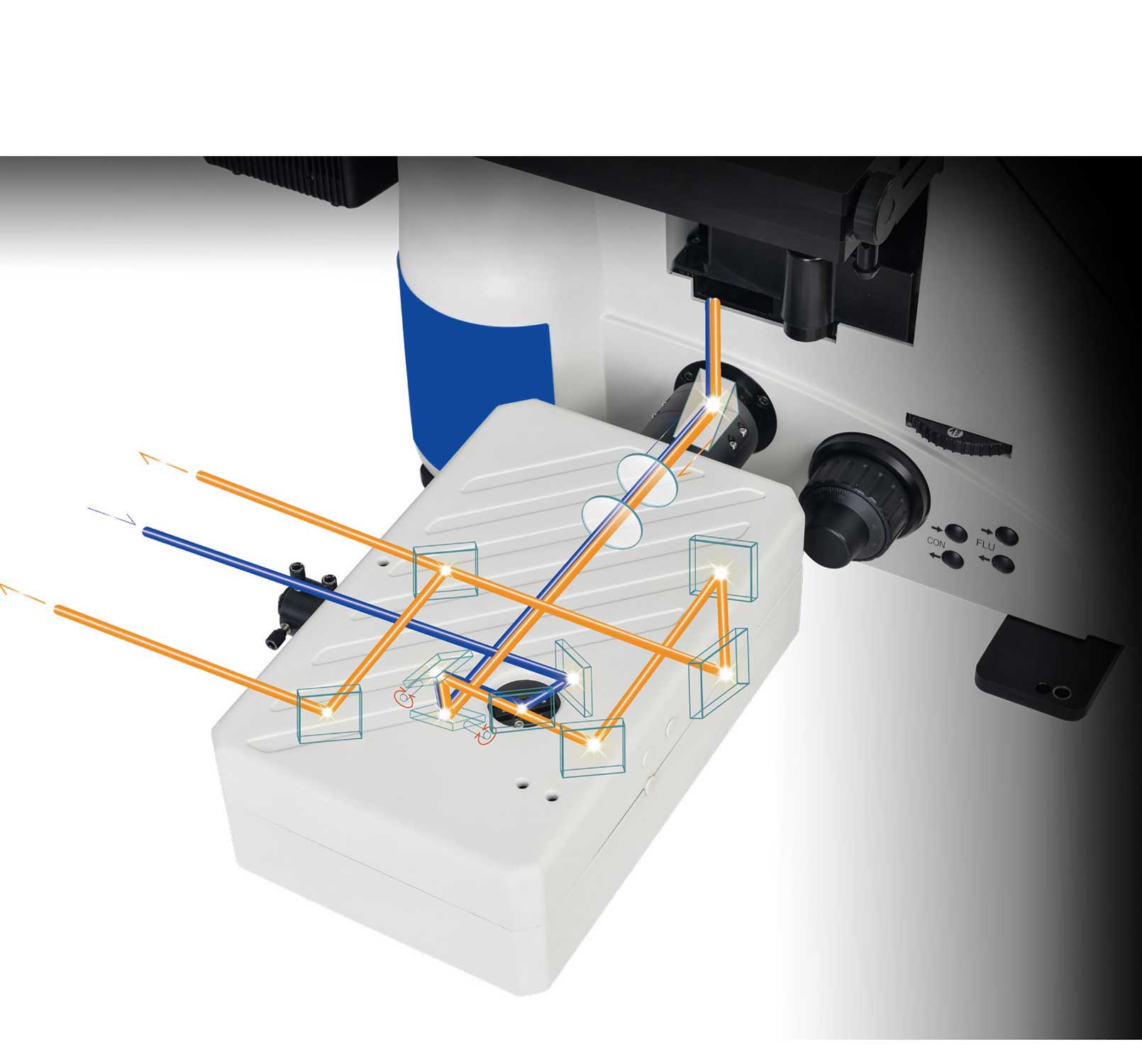
    Laser Unit RCLU Laser 405 nm,488 nm,561 nm,633/638nm, 640 nm, Motorized beam path
    Detector RCD Wavelength: 400-750nm, Detector: 4PMT
    Scanner RCS Maximum Pixel Size: 4096
    Scanning speed: 2fps(512X512), 18fps(256X256), 0.5fps(1024X1024), 0.12fps(2048X2048), 0.03fps (4096X4096)
    Scan Mode RCSM X-Y, X-Y-Z, X-Y-T
    Pinhole RCPH Hexagon shape, Continuouslv Variable Transmission(CVT)
    Confocal RCCF Field number Square Inscribed in a φ18mm Circle
    Image bit depth RCIBD 12 bits
    Compatible Microscopes RCCM Full Motorized Inverted Microscope
    Power Supply RSMPS620 Integrated SMPS based, wide voltage range, 90V-240V, 50/60Hz Supply
     
    Confocal scanning system
  • Scanning head design:High stability scan head design, connected to the detector via optical fiber for easy disassembly and handling.
  • The confocal scanning head was coupled to the left interface of the microscope body to achieve the highest quality optical path imaging.
  • The fluorescence detection channel uses no less than four independent high-sensitive detectors (PMT) and 1 transmission dic detection channel.
  • Electric hole: stevariable speed hole, adjustment range: 0 to 0.5mm
  • High sensitivity detector: The high sensitivity detector, whose quantum efficiency is QE ≥ 20%@500-550nm, which is about twice that of the traditional . Effectively improve the image quality, which is suitable for high-quality imaging of weak fluorescence. Having 2PMT in standard detector for 2 color simultaneous image acquisition and having capable to perform 4 color sequential imaging.
  • The image bit deep:16 bits
  • Pinhole: Hexagon shape, CVT. Computer controlled continuously variable single pinhole system, which should cover wider area of one Airy unit for higher brightness and without affecting sectioning performance
  • Confocal Field no. Square inscribed in ɸ18mm circle
  • Integrated design: laser, detector and signal acquisition device integrated design, placed in the same cabinet, easy to overall handling and installation.
  • Scan speed: 2 fps (512 X 512), 18 fps (256x256), 5 fps (1024x1024), 0.12 fps (2048x2048(, 0.03 fps (4096x4096), fast mode: ≥2fps (512x512); fastest: 8fps (256x256).
  • Scanning field of view: the field of vision is not less than 22mm;
  • Scan pixels: maximum 4096X4096 9 up to (8K x 8K ) output in the same field of view.
  • Optical scanning zoom:≥ 1-1000X, In order to accurately scan and locate the details, multistep.
  • Scanning mode:Any combination of points, lines, faces, x-y-z-t- λ.
  • High transmission efficiency optics for confocal
  • d. Galvano Point scanner should provide a speed of 8-10fps @ 512 x 512( without Interlacing Imaging)& max speed of 230fps @ 512 x 16.
  • System is having to possess efficient dichroic mirror with low angle incidence for better transmission efficiency .
  • Transmitted light detector will be provided for capturing bright field and DIC images
  • Laser Unit
  • The laser output of all lasers is controlled by the acoustic and optical controller (AOTF), integrated into the scanning head system, and opened with one key, to avoid the risk of color series caused by multiple channels, and to ensure the stability and accuracy of the optical path output; /direct modulation for laser attenuation and switching in synchronization with scanner.
  • Solid-state laser 405nm, Optical fiber export power≥ 20mW;
  • Solid-state laser 488nm, Optical fiber export power≥ 20mW;
  • Solid-state laser 561nm, Optical fiber export power≥ 20mW;
  • Solid-state laser 633nm/638nm, Optical fiber export power≥ 20mW;
  • Solid-state laser 640nm, Optical fiber export power≥ 20mW;
  • Software
  • Image acquisition and system automatic control function, optical road full electric control switch.
  • Complete microscope control, Scan head control and Laser control software
  • Saving of all instrument parameters along with the image for repeatable/reproducible imaging
  • Frame/line/lambda capturing, Z-Stack, Time series imaging capabilities
  • ROI bleach for FRAP experiments
  • Co-localization analysis and volume rendering
  • Multipoint analysis 
  • 6D experimental Preview
  • Multidimensional image viewer
  • Interactive movie/volume rendering
  • Macro creation
  • Segmentation
  • Volume measurement
  • Intelligent setting: according to the dye or different application requirements, the software can set the time-sharing scanning or simultaneous scanning.
  • Multidimensional microscopy imaging control: x, y, z, t and other control, to realize the automatic acquisition and processing of multi-time, multi-channel fluorescence, multipoint and z sequence.
  • Real-time multi-color image scanning and real-time multi-color image superposition, multi-dimensional (x, y, z, t) confocal image acquisition, processing, and reconstruction
  • The acquisition software has a 3 d video image reconstruction function, random space cutting, and interactive three-dimensional display. The acquisition software is the same software as the 3 D browsing software.
  • Area measurement function, can draw the partition figure of interest on the image, measure the parameters in the area, such as: area / length, etc.
  • It can recombine the acquired z-axis sequence image group, and the user can browse the sample 3 D stereo images.
  • The software has a microscope electric component control function, which can control the objective turntable rotation, fluorescent turntable rotation, condenser rotation, platform motion and z-axis focus. Can realize the slice scanning function;
  • NOMIS advanced C, Display / Image Processing / Analysis.
  • 2D / 3D / 4D analysis, Time – Lapse analysis, 3 D volume Render / orthogonal, image stitching, multi – channel color Confocal imag
  • Compatible Desktop Computer System (Workstation) Windows 10 Pro 64 bit OS English version .;
    CPU: Intel Xeon W-2225( CPU 4.0 core 4)
    RAM: 64 GB
    HDD: 1st HP Z turbo G2 512x GB, PCIe H.2 SSD.
    2nd SATA 2TB
    Optical Drive: Super Multi drive, upto x 16 speed.
    LAN: 10/100/1000network interface x 2
    Extension Slot: 2 PCI express 3.0 x 16 slots (one slot for Graphics)
    Graphics:NVIDIAQuadro RTX4000
    Monitor:32" monitors
    Suitable UPS
    Accessories RCC Cleaning cloth
      RVC Vinyl/Dust Cover
    RAK Allen keys
    RF5A Fuses
    MANUAL Instruction manual
    WARRANTY Warranty card
    Power Cord (country Specific)

    As we are continuously improving & developing our products, this websites may not be updated with advancements done. However, we try our best to update the website for latest information's
    For complete updated specifications, please do ask for latest brochures

    Contact Info